Musculoskeletal Ultrasound
The musculoskeletal system includes muscles, tendons, joints, ligaments and soft tissues. Ultrasound uses reflected high frequency sound waves to image these structures and to assess blood flow within them. Ultrasound is useful in many conditions. Common examples include;
- Shoulder (rotator cuff tears, tendonitis, bursitis, impingement syndrome)
- Elbow (collateral ligament evaluation, triceps/biceps tendons)
- Hand/wrist (carpal tunnel syndrome, tendon tears)
- Hip/knee (quadriceps/patellar tendon injuries, Baker’s cyst, bursitis)
- Ankle/foot (achilles tendon, plantar fasciitis, Morton’s neuroma, joint problems, tendon/ligament)
- Nerves and masses (tumors, cysts, etc.)
- Guided injections
What happens during the procedure
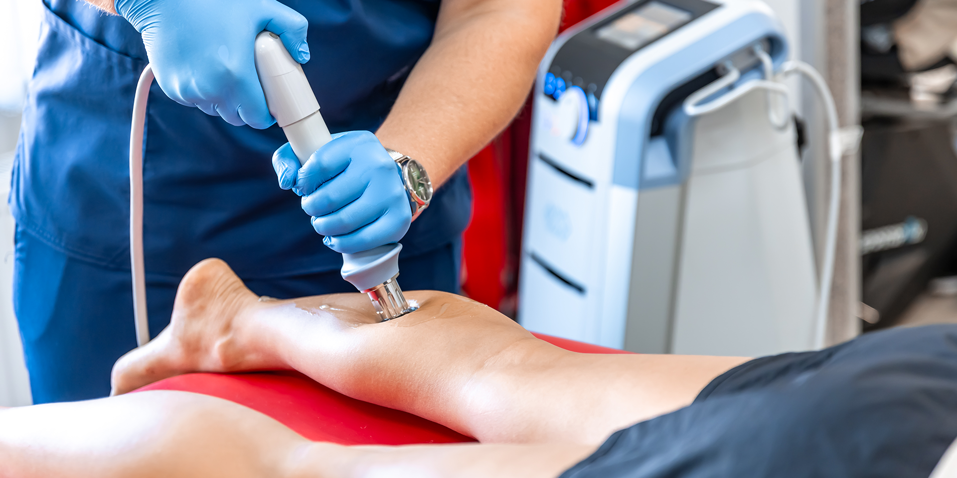
You may be asked to put on a gown. Gel is applied to the skin to improve contact between the skin and transducer (a handheld plastic probe which sends and receives the reflected sound waves that forms the image). The transducer is moved slowly over the area of interest to acquire images in different planes.
The images are displayed on a computer monitor for interpretation. Ultrasound is generally painless but discomfort from transducer pressure may be experienced if the region examined is tender.
Your images and report
A report, along with the images will be sent directly to your referring doctor. Lucan Diagnostic Imaging will store digital copies of all studies on our secure database for comparison with any future examinations.
It is important that you return to your doctor with your examination results. Whether they are normal or abnormal, your doctor needs to know promptly so that a management plan can be formulated.

© Copyright 2025 Lucan Diagnostic Imaging. All Rights Reserved. Powered by design4u.ca