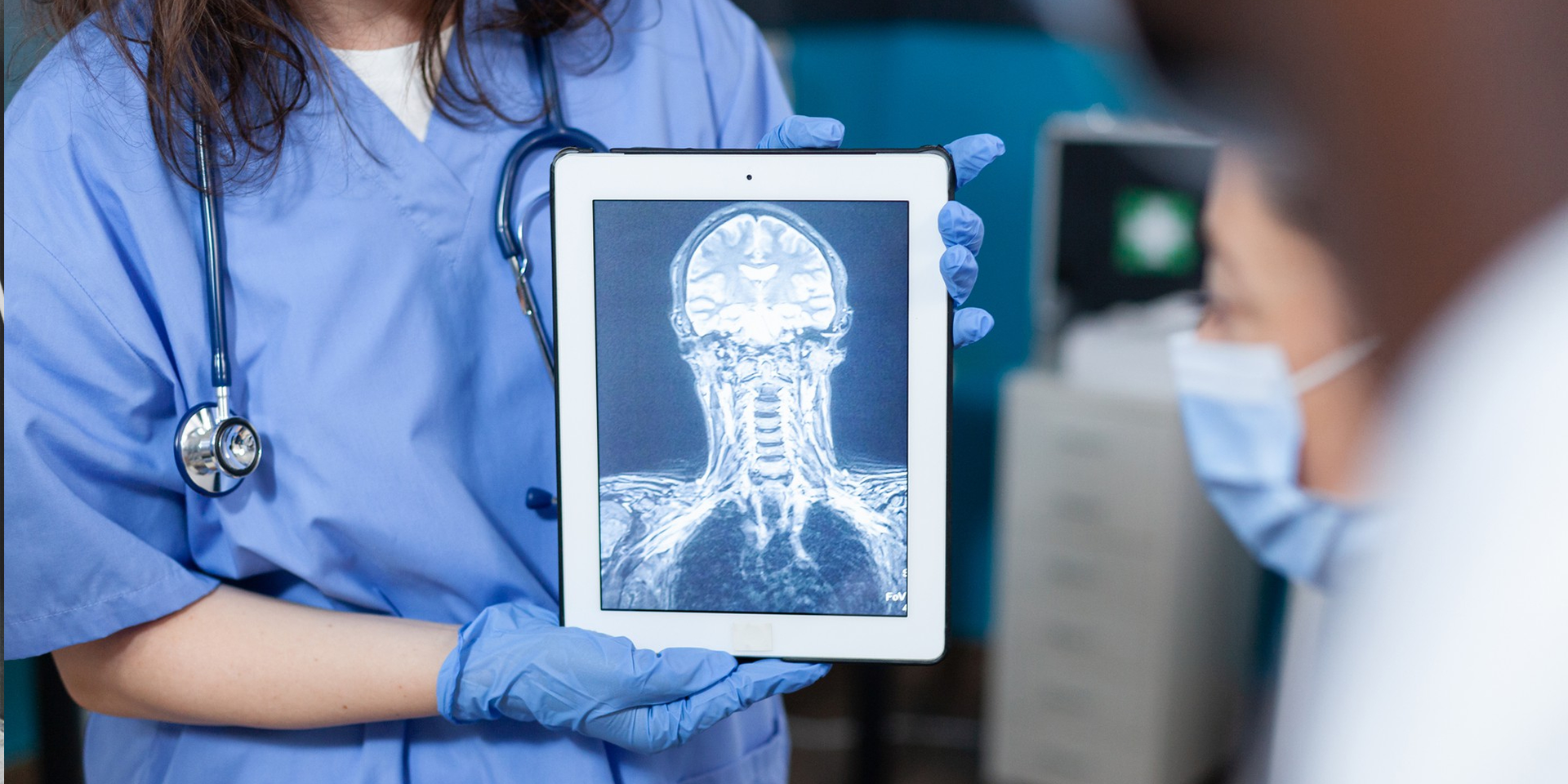
Head & Neck X-Ray
X-rays use invisible electromagnetic energy beams to make images of internal tissues, bones, and organs on film. Standard X-rays are performed for many reasons. These include diagnosing tumors or bone injuries.
X-rays are made by using external radiation to produce images of the body, its organs, and other internal structures for diagnostic purposes. X-rays pass through body tissues onto specially-treated plates (similar to camera film) and a “negative” type picture is made (the more solid a structure is, the whiter it appears on the film). Instead of film, X-rays are now typically made by using computers and digital media.
When the body undergoes X-rays, different parts of the body allow varying amounts of the X-ray beams to pass through. Images are made in degrees of light and dark. It depends on the amount of X-rays that penetrate the tissues. The soft tissues in the body (like blood, skin, fat, and muscle) allow most of the X-ray to pass through and appear dark gray on the film. A bone or a tumor, which is denser than soft tissue, allows few of the X-rays to pass through and appears white on the X-ray. At a break in a bone, the X-ray beam passes through the broken area. It appears as a dark line in the white bone.
X-rays of the spine may be performed to evaluate any area of the spine (cervical, thoracic, lumbar, sacral, or coccygeal). Other related procedures that may be used to diagnose spine, back, or neck problems include myelography (myelogram), computed tomography (CT scan), magnetic resonance imaging (MRI), or bone scans. Please see these procedures for additional information.
During the procedure
An X-ray may be performed on an outpatient basis or as part of your stay in a hospital. Procedures may vary depending on your condition and your health care provider’s practices.
Generally, an X-ray procedure of the spine, neck, or back follows this process:
- You will be asked to remove any clothing, jewelry, hairpins, eyeglasses, hearing aids, or other metal objects that may interfere with the procedure.
- If you are asked to remove any clothing, you will be given a gown to wear.
- You will be positioned on an X-ray table that carefully places the part of the spine that is to be X-rayed between the X-ray machine and a cassette containing the X-ray film or digital media. Your health care provider may also request X-ray views to be taken from a standing position.
- Body parts not being imaged may be covered with a lead apron (shield) to avoid exposure to the X-rays.
- The radiologic technologist will ask you to hold still in a certain position for a few moments while the X-ray exposure is made.
- If the X-ray is being performed to determine an injury, special care will be taken to prevent further injury. For example, a neck brace may be applied if a cervical spine fracture is suspected.
- Some spinal X-ray studies may require several different positions. Unless the technologist instructs you otherwise, it is extremely important to remain completely still while the exposure is made. Any movement may distort the image and even require another study to be done to obtain a clear image of the body part in question. You may be asked to breathe in and out during a thoracic spine X-ray.
- The X-ray beam will be focused on the area to be photographed.
- The radiologic technologist will step behind a protective window while the image is taken.
After the procedure
Generally, there is no special type of care following an X-ray of the spine, back, or neck. However, your health care provider may give you additional or alternate instructions after the procedure, depending on your particular situation.

© Copyright 2025 Lucan Diagnostic Imaging. All Rights Reserved. Powered by design4u.ca